Radiation Prevention
Is radiation harmful?
Ionizing radiation is proven to be harmful to living cells. It is capable of damaging DNA directly, which leads to cancer.
How to avoid it?
Exposure to ionizing radiation is a part of life on our planet. We all are exposed to background radiation of around 150 millirads per year. The fact that we all do not get cancer right away is a testament to the fact that our bodies repair damage done by radiation.
Even though we have repair processes, minimizing ionizing radiation exposure is wise. Most avoidable radiation nowadays comes from medical imaging, although smoking tobacco also falls into this category. Smoking a pack a day has been estimated to deliver 5000 millirads per year. The radiation comes from Polonium 210.
Ignorance
It is not pleasant, but it is worth asking medical practitioners what radiation dose is associated with the imaging process that they are going to use. They should be able to answer you if you are to give 'informed consent'. In general, the answer will not be technically acceptable, as ignorance of radiation dose and possible effects is widespread throughout society and medical organizations. If the organization has a medical physicist (they do exist), they will be able to discuss doses, but most others fluff it off. Typical answers to expect "about the same as a flight across the country". Ask for doses in millirads. Expect 3 millirads for a tooth x-ray, 30 millirads for a chest x-ray, 600 millirads per breast for x-ray mammogram, 1000 to 10,000 millirads for a CAT scan, 500 to 40,000 millirads for angiography during heart catheterization (600 millirads per minute of beam time is 'low' and the cardiologist may be scanning your heart for 40 minutes) and zero for MRI imaging. MRIs are typically very noisy, but use no ionizing radiation, whereas CAT scans are quiet, but are like getting 30 to 300 chest x-rays in one shot. For comparison, standing a mile or two from ground zero at Hiroshima exposed survivors to around 3000 millirads of radiation. Medical radiation is serious and cause for alarm.
Mammography
Mammography is breast imaging, mostly used to detect breast cancers. The majority of mammography is done by squashing a breast between two plates and then shining x-rays thru it and getting an image. The radiation dose can be as high as 600 millirads each. Wow. Two parts of this process are harmful. The squashing often is hard enough to cause swelling. It has been suggested that it is also hard enough to squeeze ductal cancer cells out of the milk ducts into other part of the breast, and maybe thru the blood to other parts of the body. Second, radiation causes cancer, and breast tissue is quite sensitive to radiation damage when compared to other tissues in the body.
What most doctors don't yet know.
MRI technology and Ultrasound are two technologies that will replace x-ray mammography. MRI uses magnetic fields and ultrasound uses sound waves. Both are much less harmful than x-rays. Recent studies show that MRI is about 3 times better at detecting cancer than x-ray mammography and ultrasound is comparable to x-ray mammography. See the image below. All images below are from a presentation made by Sentinelle Medical's president, Cameron Piron. Sentinelle Medical was dedicated to optimizing MRI technology and applications. They did some brilliant work. The company was sold to Hologic in 2010. The image below shows comparative effectiveness of MRI, X-ray, clinical breast exam, and ultrasound detection. Increased use of MRI technology would improve outcomes as it is more sensitive and does no harm.

The image below shows the difference in resolution between X-ray and MRI. The level of detail in the MRI image is better. More like a pathology slide than a shadow graph. The "O" on the right hand image is a pre-cancerous growth. Wow. Similar to dysplasia detection in a cervix and detection of polyps in a colon, detection of pre-cancerous tissue in a breast can rightly be placed in the prevention category, as cancer has not developed, but it likely will. Catching it at this stage makes life much easier for the patient and the doctor.

The image below shows the improvement in resolution of MRI imaging over the years and the time required to acquire the image.

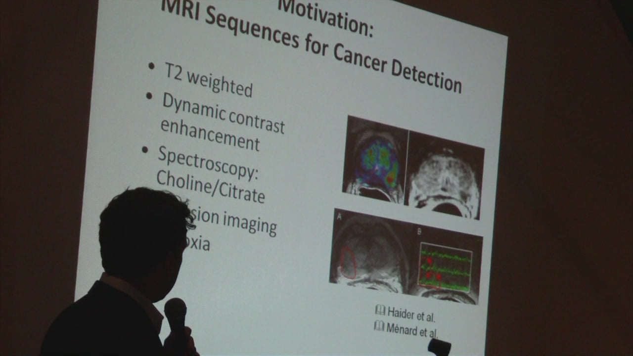
MRI is a data rich type of spectroscopy. Information on chemical species and processes within the body can be determined. In the lower left image, areas of low oxygen (hypoxia) are circled. Cancer tends to exist in low oxygen environments. These are images of prostates made by MRI, using Sentinelle accessories.
With such sensitive imaging, individualized chemotherapy monitoring can be performed. The images below show tumor size reduction over time in response to chemotherapy. Using imaging during chemotherapy allows doctors to literally see what works and make changes as needed.
